Tuesday, December 31, 2013

Allergic Contact Dermatitis

practice essentials

individuals with allergic contact dermatitis could have chronic or relapsing dermatitis, particularly if the material(s) to which they're allergic is not recognized or in the event that they practice inappropriate skin care. The longer an individual has severe dermatitis, the longer, it's believed, that the dermatitis will take to unravel once the cause is recognized.

crucial update: Allergic contact dermatitis as a result of non-latex rubber gloves

In a learn about of surgical operation personnel in Sweden with occupational allergic contact dermatitis, Pontén et al discovered proof that the situation used to be caused by 1,three-diphenylguanidine (1,3-DPG) in non-latex rubber gloves.[1] the usage of patch assessments, the investigators discovered that 12 of 16 patients reacted to 1,three-DPG. The 1,three-DPG used to be present in the gloves worn via the patients within the learn about, with the next concentration on the inside of the gloves than on the skin. In 7 of eight sufferers, contact allergic reaction to cetylpyridinium chloride was also discovered[1]

signs and symptoms

Acute allergic contact dermatitis is characterised via pruritic papules and vesicles on an erythematous base. Lichenified pruritic plaques may indicate a continual form of the situation.

individuals with allergic contact dermatitis most often boost the situation inside a couple of days of publicity, in areas that have been uncovered immediately to the allergen. certain allergens (eg, neomycin), then again, penetrate intact pores and skin poorly; in such circumstances, the onset of dermatitis could also be delayed for as much as every week following exposure.

individuals could boost well-liked dermatitis from topical medicines utilized to leg ulcers or from go-reacting systemic medicines administered intravenously.

Intraoral metallic contact hypersensitivity could end in mucositis that mimics lichen planus, which has an association with intraoral squamous cell carcinoma.

See clinical Presentation for extra element.

prognosis

Diagnostic research for allergic contact dermatitis embody the next:

Potassium hydroxide guidance and/or fungal tradition: To exclude tinea; these checks are ceaselessly indicated for dermatitis of the hands and feet Patch checking out: To identify external chemical substances to which the particular person is allergicRepeat open software test (ROAT): To determine whether a response is very important in folks that enhance weak or 1+ sure reactions to a chemical Dimethylgloxime take a look at: To determine whether a metal object comprises enough nickel to provoke allergic dermatitisSkin biopsy: could lend a hand to exclude different disorders, specifically tinea, psoriasis, and cutaneous lymphoma

See Workup for more element.

administration

The definitive treatment for allergic contact dermatitis is the identification and elimination of any attainable causal marketers; in any other case, the patient is at elevated chance for persistent or recurrent dermatitis. remedies also embrace the next:

Corticosteroids: Topical corticosteroids are the mainstay of treatment, even though acute, extreme allergic contact dermatitis, corresponding to from poison ivy, regularly must be treated with a 2-week direction of systemic corticosteroids Topical immunomodulators (TIMs): authorized for atopic dermatitis, but they're additionally prescribed for circumstances of allergic contact dermatitis once they offer security advantages over topical corticosteroids Phototherapy: Administered to individuals with persistent allergic contact dermatitis that's not managed neatly by using topical corticosteroids; these sufferers may just make the most of remedy with a combination of psoralen (a photosensitizer) and ultraviolet-A (PUVA) Immunosuppressive marketers: power immunosuppressive retailers are, in rare circumstances, used to treat recalcitrant circumstances of severe, power, standard allergic contact dermatitis or severe hand dermatitis that forestalls a affected person from working or performing day by day actions Disulfiram: occasionally, a person who is highly allergic to nickel and has extreme vesicular hand dermatitis will benefit from remedy with disulfiram (Antabuse); the drug has a chelating impact

See treatment and drugs for more element.

image libraryChronic stasis dermatitis with allergic contact de
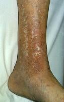
chronic stasis dermatitis with allergic contact dermatitis to quaternium-15, a preservative in moisturizer. Allergic contact dermatitis produces areas of erythema in areas of atrophie blanche and varicose veins. NextBackground

Allergic contact dermatitis (ACD) is a delayed type of caused sensitivity (allergic reaction) as a result of cutaneous contact with a selected allergen to which the affected person has developed a specific sensitivity. This allergic reaction motives inflammation of the pores and skin manifested by means of varying levels of erythema, edema, and vesiculation.

The time period contact dermatitis once in a while is used incorrectly as a synonym for allergic contact dermatitis. Contact dermatitis is inflammation of the skin precipitated by way of chemicals that right away injury the skin (see Irritant Contact Dermatitis) and by way of specific sensitivity in the case of allergic contact dermatitis.

Jadassohn first described allergic contact dermatitis in 1895. He developed the patch take a look at to establish the chemical substances to which the patient was once allergic. Sulzberger popularized patch trying out in the us in the Nineteen Thirties. The Finn chamber approach for patch trying out used to be designed within the 1970s; these chambers consist of small steel cups, typically attached to strips of tape, stuffed with allergens dispersed in either petrolatum or water. the skinny-layer fast use epicutaneous (proper) check for patch trying out turned into to be had in the united states within the Nineteen Nineties.

The importance of specific materials as reasons of allergic contact dermatitis varies with the incidence of that substance in the atmosphere. Mercury compounds as soon as have been significant causes of allergic contact dermatitis but rarely are used as topical drugs and, at the moment, are distinct as a result in of allergic contact dermatitis. Ethylenediamine, which used to be existing in the unique Mycolog cream, declined as a main result in of allergic contact dermatitis once Mycolog cream was reformulated to not contain this allergen.

a detailed history, both ahead of and after patch trying out, is a very powerful in evaluating people with allergic contact dermatitis. before patch testing, the history identifies doable causes of allergic contact dermatitis and the materials to which people are exposed that should be incorporated in patch testing. After patch checking out, the historical past determines the clinical significance of the findings. (See clinical.)

Topical corticosteroids are the mainstay of therapy, whereas a variety of symptomatic treatments can present brief-term relief of pruritus. alternatively, the definitive therapy of allergic contact dermatitis is the identification and removing of any doable causal retailers; in any other case, the patient is at increased risk for persistent or recurrent dermatitis. (See treatment.)

Go to Irritant Contact Dermatitis, Pediatric Contact Dermatitis, and Protein Contact Dermatitis for full knowledge on these issues.

PreviousNextPathophysiology

roughly 3000 chemical substances are smartly documented as specific causes of allergic contact dermatitis.

Compounds need to be less than 500 d for environment friendly penetration throughout the stratum corneum barrier, which is the water-impermeable outer layer of the skin. Small organic molecules that are chemically reactive (chemical sensitizers) bind with self-proteins to generate immunogenic neoantigens via a process termed haptenization. even supposing haptens can penetrate via intact pores and skin, sufferers with certain illness states that impair barrier perform (eg, leg ulcers, perianal dermatitis) have an increased chance of sensitization to topically utilized medicines and their vehicle elements.

Many sufferers with atopic dermatitis or allergic contact dermatitis to nickel harbor a faulty form of the filaggrin gene.[2] Filaggrin helps combination cytoskeletal proteins that form the cornified cell envelope. In its absence, the barrier is flawed.

Prehaptens are chemical substances that aren't activated through host proteins, but as an alternative require chemical transformation via oxidative derivatization by ambient or air oxidation to form hydroperoxide. Examples embody sure heady scent materials and dyes utilized in hair coloring, akin to para-phenylenediamine.

Haptens activate Toll-like receptors (TLRs) and prompt innate immunity. The importance of hapten-mediated activation of innate immunity is highlighted via the clinical remark that the irritancy of chemical substances (ie, the ability of those chemical substances to cause grossly visible skin irritation upon primary publicity) correlates with their capacity to act as contact sensitizers and to induce acute contact dermatitis.

Haptens or haptenated self-proteins are recognized through innate immune mechanisms in the skin, and this results in the elaboration of plenty of proinflammatory mediators, including interleukin (IL)–1β. because of this, pores and skin-resident dendritic cells (DCs) develop into activated. There are a couple of populations of DCs. Langerhans cells are the one DC subtype within the dermis. like every pores and skin-resident DCs, Langerhans cells efficiently accumulate antigen in the periphery and migrate to regional lymph nodes the place they existing antigen to naïve and reminiscence T cells. These DCs, which will have been directly haptenated or can have obtained haptenated proteins from their environment, migrate to skin-draining lymph nodes the place they existing peptides from haptenated proteins to set off memory and naïve T cells.

within the last step, hapten-caused irritation recruits activated effector T cells back to the preliminary website online of antigen come across in the skin. The effector T cells unencumber proinflammatory cytokines, equivalent to interferon-γ, and promote the killing of haptenated cells, resulting in the construction of the basic inflammatory rash considered in allergic contact dermatitis.

Keratinocytes are a very powerful for the advance of allergic contact dermatitis. They constitute the overwhelming majority of cells within the epidermis and form the anatomic barrier of the skin. Keratinocytes specific most TLRs, and this allows them to answer TLR4-triggering haptens, equivalent to nickel. Keratinocytes are additionally a source of IL-10, an immunosuppressive cytokine that limits the extent of contact hypersensitivity

The initial sensitization generally takes 10-14 days from initial publicity to a strong contact allergen corresponding to poison ivy. Some individuals enhance explicit sensitivity to allergens following years of chronic low-grade exposure; for example, sensitivity to chromate in cement can ultimately develop in people with power irritant contact dermatitis attributable to the alkaline nature of cement. as soon as an individual is sensitized to a chemical, allergic contact dermatitis develops within hours to a number of days of publicity.

CD4+ CCR10+ reminiscence T cells persist within the dermis after scientific decision of allergic contact dermatitis.

PreviousNextEtiology

roughly 25 chemicals look like accountable for as many as one 1/2 of all cases of allergic contact dermatitis. These embody nickel, preservatives, dyes, and fragrances.

Poison ivy

Poison ivy (Toxicodendron radicans) is the basic example of acute allergic contact dermatitis in North the united states. Allergic contact dermatitis from poison ivy is characterized by means of linear streaks of acute dermatitis that improve where plant parts had been in direct contact with the pores and skin.

Nickel

Nickel is the leading result in of allergic contact dermatitis in the world. The incidence of nickel allergic contact dermatitis in North america is rising; in distinction, new laws in Europe have resulted in a reducing incidence of nickel hypersensitivity in young and heart-aged ladies.[3, 4]

Allergic contact dermatitis to nickel generally is manifested via dermatitis on the web sites the place rings or necklaces (see the image beneath) containing nickel are worn or the place metallic objects (together with the keypads of some cell phones[5] ) containing nickel are in touch with the pores and skin.

Nickel is also regarded as a conceivable occupational allergen. employees in whom nickel is also an occupational allergen primarily include hairdressers, retail clerks, caterers, domestic cleaners, and metalworkers. people allergic to nickel every so often could advance vesicles on the sides of the fingers (dyshidrotic hand eczema or pompholyx) from nickel within the weight-reduction plan.

Allergic contact dermatitis to nickel in a necklac
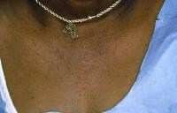
Allergic contact dermatitis to nickel in a necklace. Rubber gloves

allergic reaction to 1 or more chemicals in rubber gloves is recommended in any individual with continual hand dermatitis who wears them, except patch checking out demonstrates otherwise. Allergic contact dermatitis to chemical compounds in rubber gloves typically happens maximally on the dorsal elements of the hand. frequently, a cutoff of dermatitis happens on the forearms where skin is no longer in contact with the gloves. individuals allergic to chemical compounds in rubber gloves could improve dermatitis from different exposures to the chemical compounds (eg, under elastic waistbands).

Hair dye and transient tattoos

p-Phenylenediamine (PPD) is a typical section of and sensitizer in everlasting hair dye products and brief henna tattoos[6] ; publicity in to it in hair dye products could result in acute dermatitis with extreme facial edema. severe local reactions from PPD may just occur in black henna tattoos in adults and children. Epidemiologic information point out that the median occurrence of certain patch check reactions to PPD among dermatitis patients is four.three% (growing) in Asia, four% (plateau) in Europe, and 6.2% (decreasing) in North the us.[7]

Textiles

people allergic to dyes and permanent press and wash-and-wear chemicals delivered to textiles typically advance dermatitis on the trunk, which occurs maximally on the lateral sides of the trunk but spares the vault of the axillae. main lesions could also be small follicular papules or may be in depth plaques.

individuals in whom this allergic contact dermatitis is suspected must be examined with a collection of fabric chemicals, particularly if movements patch testing reveals no allergic reaction to formaldehyde. New garb is perhaps to provoke allergic contact dermatitis, for the reason that most allergens decrease in concentration in apparel following repeated washings.

Preservatives

Preservative chemical compounds introduced to cosmetics, moisturizers, and topical medications are main reasons of allergic contact dermatitis (see the picture below). the chance of allergic contact dermatitis appears to be best possible to quaternium-15, followed by way of allergic contact dermatitis to isothiazolinones. Kathon CG is methylchloroisothiazolinone in combination with methylisothiazolinone.

Methylisothiazolinone is now used as an individual preservative and could also be a significant allergen.[8]

even though parabens are among the many most generally used preservatives, they don't seem to be a established cause of allergic contact dermatitis.

Severe allergic contact dermatitis resulting from 
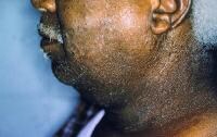
extreme allergic contact dermatitis as a result of preservatives in sunscreen. Patch testing was terrible to the active ingredients within the sunscreen.

Schnuch et al estimated that preservatives present in leave-on topical products varied over 2 orders of magnitude in relative sensitization risk.[9]

Formaldehyde is an incredible result in of allergic contact dermatitis (see the picture beneath). sure preservative chemicals widely utilized in shampoos, lotions, other moisturizers, and cosmetics are termed formaldehyde releasers (ie, quaternium-15 [Dowicil 200], imidazolidinyl urea [Germall 115], and isothiazolinones[9] ).

Onycholysis developing from allergic contact derma
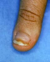
Onycholysis creating from allergic contact dermatitis to formaldehyde used to harden nails. Fragrances

people may just increase allergy to fragrances. Fragrances are discovered not handiest in perfumes, colognes, aftershaves, deodorants, and soaps, but additionally in a lot of different products, ceaselessly as a masks to camouflage an unpleasant smell. Unscented merchandise could contain fragrance chemical substances used as a portion of the product and no longer labeled as scent.

folks allergic to fragrances should use scent-free products. sadly, the exact chemical compounds answerable for a scent in a product aren't labeled. 4 thousand totally different scent molecules are to be had to formulate perfumes. The fragrance business shouldn't be required to free up the names of elements used to compose a fragrance in the U.S., even when people boost allergic contact dermatitis to fragrances found in topical medications.

Deodorants is also the most common cause of allergic contact dermatitis to fragrances as a result of they are utilized to occlude pores and skin that is frequently abraded by way of shaving in girls.

massage and bodily therapists and geriatric nurses are at greater risk of occupational allergic contact dermatitis to fragrances.

Corticosteroids

within the final decade, it has develop into clear that some individuals with persistent dermatitis strengthen hypersensitive reaction to topical corticosteroids. Most affected people may also be treated with some topical corticosteroids, however an individual can also be allergic to all topical and systemic corticosteroids. Budesonide and tixocortol pivalate are useful patch take a look at corticosteroids for making a choice on individuals allergic to topical corticosteroids.

Neomycin

the danger of hypersensitive reaction to neomycin is said instantly to the extent of its use in a inhabitants. the risk of allergy to neomycin is way larger when it is used to treat continual stasis dermatitis and venous ulcers than when it is used as a topical antibiotic on cuts and abrasions in children. think that people allergic to neomycin are allergic to chemically related aminoglycoside antibiotics (eg, gentamicin, tobramycin).[10] avoid these medicine each topically and systemically in folks allergic to neomycin.

Benzocaine

steer clear of topical use of benzocaine. Benzocaine is integrated in most same old patch check trays. people allergic to benzocaine may safely use or be injected with lidocaine (Xylocaine), which does not move-react with benzocaine.

a lot of people complain of adversarial reactions to sunscreens, but many of those people are no longer allergic to the sunscreen supplies. They is also allergic to preservatives in these products or can have nonspecific cutaneous inflammation from these products.

Photoallergy

now and again, folks strengthen photoallergic contact dermatitis. Allergic contact dermatitis is also accentuated by ultraviolet (UV) gentle, or sufferers may just improve an allergy handiest when a chemical is present on the pores and skin and when the skin is exposed sufficiently to ultraviolet light A (UV-A; 320-four hundred nm).

PreviousNextEpidemiologyUnited States data

The national well being and nutrition Examination Survey (NHANES) estimated the incidence of contact dermatitis to be 13.6 cases per a thousand inhabitants, the usage of bodily examinations by means of dermatologists of a specific pattern of sufferers. NHANES underreported the prevalence when put next with the physical examination findings.

The nationwide Ambulatory hospital therapy Survey performed in 1995 estimated eight.4 million outpatient visits to American physicians for contact dermatitis. This was the second most common dermatologic diagnosis. Of place of job visits to dermatologists, 9% are for dermatitis. At a student health middle dermatology health facility, 3.1% of sufferers introduced for allergic contact dermatitis, and a couple of.three% introduced for irritant contact dermatitis.

The authentic check site can present accurate normal knowledge on widespread allergens. The Contact Allergen administration program is equipped as a service to the American Contact Dermatitis Society (ACDS) contributors and is especially precious for allergens present in topical skin care products. The Contact Allergen substitute Database (CARD) incorporates more than 8100 known ingredients cataloged in additional than 5500 commercial skincare merchandise and is available as a Smartphone software.

international records

A Swedish study discovered that incidence of allergic contact dermatitis of the fingers used to be 2.7 circumstances per a thousand population. A Dutch learn about discovered that occurrence of allergic contact dermatitis of the arms was 12 instances per one thousand inhabitants.

Race, sex, and age-related demographics

No racial predilection exists for allergic contact dermatitis. Allergic contact dermatitis is extra fashionable in ladies than in males. This predominantly is as a result allergic reaction to nickel, which is rather more well-liked in women than in men in most nations.

Allergic contact dermatitis may just happen in neonates. In elderly individuals, the improvement of allergic contact dermatitis could also be delayed rather, but the dermatitis may be more continual as soon as developed. Contact hypersensitivity to topical medicaments is extra in style in individuals older than 70 years.[11]

PreviousNextPrognosis

individuals with allergic contact dermatitis may have power or relapsing dermatitis, in particular if the material(s) to which they are allergic shouldn't be recognized or in the event that they proceed to follow skin care that's not appropriate (ie, they proceed to make use of harsh chemical compounds to wash their pores and skin, they do not observe creams with ceramides or bland emollients to protect their skin).

The longer a person has severe dermatitis, the longer it's believed it's going to take the dermatitis to unravel as soon as the lead to is identified.

Some folks have power dermatitis following allergic contact dermatitis, which seems to be authentic especially in folks allergic to chrome.

a selected problem is neurodermatitis (lichen simplex chronicus), wherein individuals many times rub or scratch a neighborhood initially affected by allergic contact dermatitis.

Mortality

death from allergic contact dermatitis is unusual in the united states. Allergic contact dermatitis to the weed wild feverfew brought about deaths in India when the seeds contaminated wheat shipments to India. This plant then become in style and a first-rate lead to of severe airborne allergic contact dermatitis.

PreviousNextPatient schooling

patients have one of the best prognosis when they are able to understand that the materials to which they are allergic and the best way to steer clear of further exposures. present sufferers with as so much knowledge as possible regarding the chemical to which they're allergic, including all recognized names of the chemical. internet sites, Smartphone applications, same old textbooks, and the real test kit incorporate normal details about the chemicals.

vulnerable people want to learn the record of ingredients earlier than applying cosmetic merchandise to their pores and skin, on account that preservative chemical compounds are used extensively in client, medical, and place of work merchandise. the same chemical can have totally different names when used for consumer or industrial functions.

provide pamphlets with coloration footage of poison ivy to individuals allergic to the plant. The American Academy of Dermatology also has pamphlets on allergic contact dermatitis and hand eczema.

For affected person schooling knowledge, see the pores and skin, Hair, and Nails center, as well as Contact Dermatitis.

PreviousProceed to medical Presentation , Allergic Contact Dermatitis

Drug Eruptions

observe essentials

Drug eruptions can mimic a variety of dermatoses. The morphologies are myriad and embrace morbilliform, urticarial, papulosquamous, pustular, and bullous. drugs may additionally lead to pruritus and dysesthesia without an obtrusive eruption. A drug-induced reaction will have to be thought to be in any patient who's taking medicines and who all at once develops a symmetric cutaneous eruption.

crucial replace: Drug patch exams can identify agents liable for severe cutaneous adversarial reactions

In a find out about of 134 patients with severe cutaneous adversarial drug reactions, Barbaud et al found that drug patch tests will be safely used to identify the accountable drug. Of the 134 patients, forty five had acute generalized exanthematous pustulosis (AGEP), 72 had drug reaction with eosinophilia and systemic signs (dress), and 17 had Stevens-Johnson syndrome (SJS)/toxic epidermal necrolysis (TEN). sure drug patch take a look at results were bought for sixty four% of the sufferers within the dress crew, 58% of these within the AGEP workforce, and 24% of these in the SJS/TEN crew; 1 AGEP affected person experienced relapse.[1]

indicators and symptoms

the first steps within the historical past are as follows:

evaluation the patient’s complete medicine checklist, together with prescription and over-the-counter drugsDocument any historical past of earlier hostile reactions to medicine or foodsConsider different etiologies (eg, viral exanthems and bacterial infections)notice any concurrent infections, metabolic problems, or immunocompromise

in addition, the following should be mentioned and unique:

Interval between introduction of a drug and onset of the eruptionRoute, dose, period, and frequency of drug administrationUse of parenterally administered medicine (extra more likely to cause anaphylaxis)Use of topically utilized drugs (more prone to result in delayed-kind hypersensitivity)Use of a couple of courses of remedy and extended administration (risk of allergic sensitization)Any improvement after drug withdrawal and any response with readministration

physical examination should handle medical options that can point out a extreme, probably existence-threatening drug response, together with the next:

Mucous membrane erosionsBlistersNikolsky signConfluent erythemaAngioedema and tongue swellingPalpable purpuraSkin necrosisLymphadenopathyHigh fever, dyspnea, or hypotension

it is important to savor the morphology and bodily options of drug eruptions, as follows:

AcneiformAcral erythema (erythrodysesthesia)AGEPDermatomyositislikeDRESSErythema multiforme (EM), together with EM minor, SJS, TEN, and SJS/TEN overlapErythema nodosumErythrodermaFixed drug eruptionsHypersensitivity syndromeLeukocytoclastic vasculitisLichenoidLupusMorbilliform or exanthematousPseudoporphyria[2] Serum sickness and serum sickness–likeSweet syndrome (acute febrile neutrophilic dermatosis)UrticariaVesiculobullous

See clinical Presentation for extra detail.

diagnosis

With gentle asymptomatic eruptions, the history and bodily examination are steadily enough for prognosis; with extreme or persistent eruptions, additional diagnostic testing could also be required, as follows:

BiopsyComplete blood rely (CBC) with differentialSerum chemistry research (particularly for electrolyte balance and indices of renal or hepatic operate in patients with extreme reactions) Antibody or immunoserology testsDirect cultures to research a major infectious etiology or secondary infectionUrinalysis, stool guaiac assessments, and chest radiography for vasculitisSkin prick or patch testing to substantiate the causative agent

See Workup for more detail.

management

rules of medical care are as follows:

the final word purpose is to identify and discontinue the offending medication if possiblePatients can on occasion continue to be treated thru morbilliform eruptions; nevertheless, all sufferers with extreme morbilliform eruptions must be monitored for mucous membrane lesions, blistering, and pores and skin sloughing remedy of a drug eruption is determined by the particular kind of reactionTherapy for exanthematous drug eruptions is supportive, involving the administration of oral antihistamines, topical steroids, and moisturizing lotions extreme reactions (eg, SJS, TEN, and hypersensitivity reactions) warrant medical institution admissionTEN is very best managed in a burn unit, and intravenous immunoglobulin (IVIG) may make stronger effects[3, 4, 5] Hypersensitivity syndrome, may just have to be treated with liver transplantation if the offending drug shouldn't be stopped in time; remedy with systemic corticosteroids has been recommended

for most drug eruptions, full restoration without any complications is expected; however, the next must be mentioned:

sufferers with exanthematous eruptions should expect mild desquamation because the rash resolvesPatients with hypersensitivity syndrome are vulnerable to changing into hypothyroid, on a regular basis inside the first four-12 weeks after the response The prognosis for sufferers with TEN is guarded; scarring, blindness, and death are that you can think of

See therapy and medication for extra element.

image libraryStevens-Johnson syndrome. Stevens-Johnson syndrome. NextBackground

Drug eruptions can mimic a wide range of dermatoses. The morphologies are myriad and embrace morbilliform (most common, see picture under), urticarial, papulosquamous, pustular, and bullous. medicines can also cause pruritus and dysesthesia with out an obtrusive eruption.

Morbilliform drug eruption. Morbilliform drug eruption.

A drug-brought about reaction will have to be regarded as in any patient who's taking medicines and who suddenly develops a symmetric cutaneous eruption. drugs which can be known for inflicting cutaneous reactions embody antimicrobial sellers,[6] nonsteroidal anti-inflammatory drugs (NSAIDs), cytokines, chemotherapeutic sellers, anticonvulsants, and psychotropic marketers.

urged identification and withdrawal of the offending agent could assist limit the toxic effects associated with the drug. the decision to discontinue a potentially crucial drug frequently presents a dilemma.

PreviousNextPathophysiology

Drug eruptions may be divided into immunologically and nonimmunologically mediated reactions.

Immunologically mediated reactions

Coombs and Gell proposed four sorts of immunologically mediated reactions, as follows:

type I is immunoglobulin E (IgE)–established reactions, which lead to urticaria, angioedema, and anaphylaxis (see the picture under). Urticaria. Urticaria. kind II is cytotoxic reactions, which result in hemolysis and purpura (see the picture beneath).Oral ulcerations in a patient receiving cytotoxic 
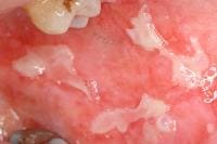
Oral ulcerations in a affected person receiving cytotoxic remedy. sort III is immune advanced reactions, which lead to vasculitis, serum sickness, and urticaria.sort IV is delayed-kind reactions with cell-mediated hypersensitivity, which result in contact dermatitis, exanthematous reactions, and photoallergic reactions.

Insulin and different proteins are related to kind I reactions. Penicillin, cephalosporins, sulfonamides, and rifampin are known to result in type II reactions. Quinine, salicylates, chlorpromazine, and sulfonamides can cause type III reactions. kind IV reactions, the most typical mechanism of drug eruptions, are steadily encountered in instances of contact hypersensitivity to topical medicines, such as neomycin. Sulfonamides are most ceaselessly related to poisonous epidermal necrolysis (TEN).

even though most drug eruptions are type IV hypersensitivity reactions, best a minority are IgE-dependent. that's, antibodies can be confirmed in less than 5% of cutaneous drug reactions. type IV cell-mediated reactions usually are not dose established, they usually commence 7-20 days after the remedy is began, they are going to involve blood or tissue eosinophilia, and so they may recur if medicine chemically associated to the causative agent are administered.

Nonimmunologically mediated reactions

Nonimmunologically mediated reactions may be categorised in keeping with the following features: accumulation, adversarial results, direct unlock of mast cell mediators, idiosyncratic reactions, intolerance, Jarisch-Herxheimer phenomenon, overdosage, or phototoxic dermatitis. (signs of Jarisch-Herxheimer reactions disappear with persevered therapy. Drug remedy must be persisted except the an infection is fully eradicated.)

An example of accumulation is argyria (blue-gray discoloration of pores and skin and nails) seen with use of silver nitrate nasal sprays.

adversarial effects are normal however negative effects of a drug. for instance, antimetabolite chemotherapeutic marketers, reminiscent of cyclophosphamide, are related to hair loss.

The direct unlock of mast cell mediators is a dose-based phenomenon that doesn't involve antibodies. for instance, aspirin and different NSAIDs lead to a shift in leukotriene manufacturing, which triggers the discharge of histamine and different mast-cell mediators. Radiographic contrast material, alcohol, cytokines, opiates, cimetidine, quinine, hydralazine, atropine, vancomycin, and tubocurarine also may just lead to release of mast-cell mediators.

Idiosyncratic reactions are unpredictable and now not explained by the pharmacologic residences of the drug. An instance is the individual with infectious mononucleosis who develops a rash when given ampicillin.

Imbalance of endogenous vegetation may happen when antimicrobial dealers preferentially suppress the growth of 1 species of microbe, allowing other species to develop vigorously. for example, candidiasis often happens with antibiotic therapy.

Intolerance may occur in sufferers with altered metabolism. as an instance, folks who are slow acetylators of the enzyme N -acetyltransferase are extra doubtless than others to develop drug-prompted lupus in line with procainamide.

Jarisch-Herxheimer phenomenon is a response because of bacterial endotoxins and microbial antigens which are liberated via the destruction of microorganisms. The response is characterised by means of fever, delicate lymphadenopathy, arthralgias, transient macular or urticarial eruptions, and exacerbation of preexisting cutaneous lesions. The reaction just isn't a sign to prevent therapy as a result of signs unravel with endured treatment. This response can also be viewed with penicillin treatment for syphilis, griseofulvin or ketoconazole therapy for dermatophyte infections, and diethylcarbamazine remedy for oncocerciasis.

Overdosage is an exaggerated response to an increased quantity of a drugs. as an instance, increased doses of anticoagulants may just lead to purpura.

Phototoxic dermatitis is an exaggerated sunburn response because of the formation of poisonous photoproducts, reminiscent of free radicals or reactive oxygen species (see the image under).

Phototoxic reaction after use of a tanning booth. 
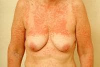
Phototoxic reaction after use of a tanning sales space. be aware sharp cutoff the place garb blocked publicity. PreviousNextFrequencyUnited States

Drug eruptions occur in approximately 2-5% of inpatients and in higher than 1% of outpatients.

world

Drug eruptions happen in roughly 2-3% of inpatients.

PreviousNextMortality/Morbidity

Most drug eruptions are delicate, self-restricted, and usually unravel after the offending agent has been discontinued. severe and doubtlessly life-threatening eruptions happen in roughly 1 in 1000 hospital patients. Mortality charges for erythema multiforme (EM) major are considerably better. Stevens-Johnson syndrome (SJS) has a mortality rate of not up to 5%, whereas the rate for TEN techniques 20-30%; most sufferers die from sepsis.

sex

opposed cutaneous reactions to medication are more everyday in women than in males.

Age

elderly patients have an increased incidence of hostile drug reactions.

PreviousProceed to medical Presentation , Drug Eruptions

Monday, December 30, 2013

Nummular Dermatitis

heritage

Nummular (that means "coin-formed") dermatitis is a form of eczema. Nummular dermatitis is characterised by round-to-oval erythematous plaques most commonly discovered on the legs and arms. Lesions steadily start as papules, which then coalesce into plaques with scale. Early nummular dermatitis lesions is also studded with vesicles containing serous exudate. they're regularly very pruritic.

NextPathophysiology

Nummular dermatitis is a condition restricted to the skin. It has not too long ago been categorised as a form of atopic dermatitis.

Little is known in regards to the pathophysiology of nummular dermatitis, but it's incessantly accompanied by means of xerosis. Dryness of the skin ends up in dysfunction of the epidermal lipid barrier; this may occasionally permit permeation of environmental allergens, which result in an allergic or irritant response.[1, 2] that is supported by one study that showed that elderly patients with nummular dermatitis had elevated sensitivity to environmental aeroallergens when compared with age-matched controls. This impaired cutaneous barrier in the environment of nummular dermatitis may also result in increased susceptibility to allergic contact dermatitis to supplies comparable to metals.[3]

Onset has been related to drugs. Onset of severe, generalized nummular lesions has been said in affiliation with interferon and ribavirin remedy for hepatitis C.[4, 5] affiliation with use of inhibitors of tumor necrosis factor has also been mentioned.[6]

Onset has also been described in association with mercury in dental amalgams. Hypersensitivity to the metals within the mouth is posulated to be adequate to force an immune response that ends up in cutaneous nummular plaques.

because of the intense pruritus associated with nummular dermatitis, the potential function of mast cells in the illness process has been investigated. increased numbers of mast cells have been observed in lesional when put next with nonlesional samples in individuals with nummular dermatitis.

One learn about identified neurogenic contributors to irritation in each nummular dermatitis and atopic dermatitis by using investigating the association between mast cells and sensory nerves and identifying the distribution of neuropeptides in the dermis and higher dermis of patients with nummular eczema. Researchers hypothesized that unlock of histamine and different inflammatory mediators from mast cells could initiate pruritus by using interacting with neural C-fibers. The research showed that dermal contacts between mast cells and nerves had been elevated in quantity in each lesional and nonlesional samples of nummular eczema when put next with commonplace controls. in addition, substance P and calcitonin gene-related peptide fibers had been prominently increased in lesional samples when put next with nonlesional samples from sufferers with nummular eczema. These neuropeptides may stimulate free up of other cytokines and promote irritation.[7, 8, 9]

other analysis has validated that mast cells existing within the dermis of sufferers with nummular eczema may have lowered chymase job, imparting decreased ability to degrade neuropeptides and protein. This dysregulation might result in reduced capability of the enzyme to suppress inflammation.

PreviousNextEpidemiologyFrequencyUnited States

The occurrence of nummular dermatitis is 2 instances per 1000 folks. Dermatitis (eg, atopic, asteatotic, dyshidrotic, nummular, hand) is likely one of the most common dermatologic stipulations.

international

The incidence internationally is the same as it is in the united states.

Mortality/MorbidityPruritus, steadily worst at night time, may just lead to irritability, insomnia, or both.Secondary infection could result in lesions that ooze serosanguineous exudate. the commonest organism published by means of culture is Staphylococcus aureus.Generalized flares may just require mattress rest, oral antibiotics, a groovy atmosphere, systemic antibiotics, and/or systemic steroids.increased contact sensitivity to environmental antigens (especially metals) could limit means to tolerate those antigens, particularly apparel, steel snaps, jewellery, dental amalgams or occupational publicity. Race

No racial predilection has been observed for nummular dermatitis.

sex

Nummular dermatitis is more common in males than in women (see Age below).

Age

Nummular dermatitis has 2 peaks of age distribution. the most typical is within the sixth to seventh decade of life. this is most continuously seen in men. A smaller top occurs within the 2nd to third decade of lifestyles, which is most regularly seen in affiliation with atopic dermatitis. that is more ceaselessly viewed in ladies. it is exclusive in kids.

PreviousProceed to clinical Presentation , Nummular Dermatitis

Drug-prompted Pigmentation

history

opposed cutaneous reactions to drugs are a typical reason for consultations with dermatologists. Drug-brought about pores and skin disorders could occur in a number of methods. drugs may cause exanthems, urticaria, hypersensitivity syndromes, pustular eruptions, erythema multiforme, poisonous epidermal necrolysis, cutaneous necrosis, and strange pigmentation of the pores and skin and mucosa. even though pigmentary modifications caused by medication frequently end in a limited level of morbidity, these adjustments is also very irritating to the patient.

The picture beneath depicts a patient with amiodarone pigmentation.

Amiodarone pigmentation. Amiodarone pigmentation.

Drug-triggered pigmentary abnormalities is also classified into 3 teams, which can be (1) hyperpigmentation/melanosis, (2) hypopigmentation/leukoderma, and (3) dyspigmentation or prevalence of odd skin colour.

A related article is fastened Drug Eruptions. moreover, the Medscape opposed Drug event Reporting useful resource heart may be of hobby.

NextPathophysiology

a couple of pathologic mechanisms are liable for drug-triggered pigmentation problems. when compared with the immunological etiology underlying many drug allergies, most circumstances of pharmacologic pigmentation should not immunologically mediated.

The pathogenesis underlying drug-related dyspigmentation can be classified into 3 mechanisms, which can be (1) drug or drug metabolite deposition in the dermis and epidermis, (2) improved melanin manufacturing with or with out an increase within the selection of active melanocytes, and (three) drug-caused postinflammatory modifications to pores and skin. in a similar fashion, chemical hypopigmentation can be idea to occur thru a lot of pathologic mechanisms, together with a reduced number of skin melanocytes, enzymatic blockade of melanogenesis, and inhibition of melanosome transfer.

PreviousNextEpidemiologyFrequencyUnited States

the rate of drug-induced dyspigmentation varies relying on the drug and cumulative dose. Some medicine, similar to amiodarone, have been stated to have a price of blue-gray dyspigmentation as excessive as 24% when the cumulative dose is larger than 200 mg.

international

Drug-induced skin pigmentation is estimated to account for 10-20% of all cases of got dyspigmentation global.

Mortality/Morbidity

Drug-brought on pigmentation isn't generally related to elevated mortality or morbidity, even if it will end in substantial psychological and social impairment.

Race

Drug-precipitated pigmentary modifications can occur in individuals of any race, however hypomelanosis is viewed more ceaselessly and appears extra dramatically in patients with darker-pigmented skin. moreover, individuals with darker pores and skin ceaselessly exhibit more excessive hyperpigmentation than folks with honest pores and skin.[1]

sex

No variations are suggested within the incidence of drug-related pigmentation amongst adult males versus women.

Age

Drug-related dyspigmentation is seen in persons of all a while.

PreviousProceed to clinical Presentation , Drug-triggered Pigmentation

Sunday, December 29, 2013

Bronchitis

follow necessities

Bronchitis is characterised by inflammation of the bronchial tubes (bronchi), the air passages that stretch from the trachea into the small airways and alveoli. It is among the prime stipulations for which sufferers seek medical care.

essential update: Overprescription of antibiotics for sore throats and bronchitis nonetheless well-liked

Two studies, the results of which were offered at IDWeek on October three, 2013, found that antibiotics are nonetheless a great deal overprescribed for sore throats and bronchitis.[1, 2]

In an analysis of data on visits to main care physicians and emergency departments for sore throats and acute bronchitis between the late Nineteen Nineties and 2010 from the national Ambulatory hospital therapy Survey and the nationwide clinic Ambulatory medical care Survey, docs prescribed antibiotics at seventy three% of visits for acute bronchitis and 60% of visits for sore throats.[1, 2] on the other hand, the prescribing charge should be zero% for acute bronchitis and 10% for sore throats.

indicators and signs

a complete history must be acquired, together with knowledge on publicity to poisonous elements and smoking. symptoms of bronchitis embrace the next:

Cough (probably the most repeatedly observed symptom)Sputum production (clear, yellow, green, or even blood-tinged)Fever (reasonably bizarre; in conjunction with cough, suggestive of influenza or pneumonia)Nausea, vomiting, and diarrhea (rare)normal malaise and chest pain (in severe instances)Dyspnea and cyanosis (most effective viewed with underlying chronic obstructive pulmonary illness [COPD] or some other situation that impairs lung operate) Sore throatRunny or stuffy noseHeadacheMuscle achesExtreme fatigue

physical examination findings in acute bronchitis are variable and may just embody the following:

Diffuse wheezes, excessive-pitched steady sounds, and the usage of accent muscle tissue (in extreme circumstances)Diffuse diminution of air consumption or inspiratory stridor (indicative of bronchial or tracheal obstruction)Sustained heave along the left sternal border (indicative of proper ventricular hypertrophy secondary to continual bronchitis)Clubbing on the digits and peripheral cyanosis (indicative of cystic fibrosis)Bullous myringitis (suggestive of mycoplasmal pneumonia)Conjunctivitis, adenopathy, and rhinorrhea (suggestive of adenoviral an infection)

See Presentation for more element.

analysis

Bronchitis may be suspected in sufferers with an acute respiratory infection with cough; but, because many more critical illnesses of the lower respiratory tract cause cough, bronchitis should be regarded as a prognosis of exclusion.

research that may be helpful embrace the next:

full blood depend (CBC) with differentialProcalcitonin ranges (to tell apart bacterial from nonbacterial infections)Sputum cytology (if the cough is chronic)Blood tradition (if bacterial superinfection is suspected)Chest radiography (if the affected person is elderly or bodily findings counsel pneumonia)Bronchoscopy (to exclude foreign physique aspiration, tuberculosis, tumors, and different power illnesses)Influenza testsSpirometryLaryngoscopy (to exclude epiglottitis)

See Workup for more element.

management

remedy is in most cases excited about alleviation of signs. handle acute bronchitis is essentially supportive. deal with continual bronchitis comprises avoidance of environmental irritants.

sellers employed for symptomatic remedy embody the following:

crucial cough suppressants (eg, codeine and dextromethorphan) – quick-time period symptomatic reduction of coughing in acute and persistent bronchitis short-acting beta-agonists (eg, ipratropium bromide and theophylline) – keep watch over of bronchospasm, dyspnea, and power cough in steady sufferers with chronic bronchitis; a long-acting beta-agonist plus an inhaled corticosteroid can also be provided to keep an eye on continual cough Nonsteroidal anti-inflammatory drugs (NSAIDs) – remedy of constitutional symptoms of acute bronchitis, including gentle-to-reasonable ache Antitussives/expectorants (eg, guaifenesin) – remedy of cough, dyspnea, and wheezingMucolytics – administration of reasonable-to-extreme COPD, particularly in iciness

amongst otherwise wholesome individuals, antibiotics have not validated any consistent advantage in acute bronchitis. the next recommendations had been made with admire to remedy of acute bronchitis with antibiotics:

Acute bronchitis must no longer be treated with antibiotics unless comorbid prerequisites pose a risk of great complicationsAntibiotic remedy is really useful in aged (>sixty five years) patients with acute cough if they have got had a hospitalization previously yr, have diabetes mellitus or congestive coronary heart failure, or are receiving steroids Antibiotic treatment is really useful in patients with acute exacerbations of continual bronchitis

In stable patients with chronic bronchitis, long-term prophylactic remedy with antibiotics is not indicated.

Influenza vaccination may cut back the incidence of upper respiratory tract infections and, due to this fact, reduce the incidence of acute bacterial bronchitis. it is usually less efficient in preventing illness than in fighting severe problems and death.

See remedy and drugs for extra element.

NextBackground

Bronchitis is likely one of the high prerequisites for which patients are searching for hospital treatment. it is characterised by using irritation of the bronchial tubes (or bronchi), the air passages that stretch from the trachea into the small airways and alveoli. (See medical Presentation.)

power bronchitis is defined clinically as cough with sputum expectoration for a minimum of 3 months a year all through a period of 2 consecutive years. persistent bronchitis is related to hypertrophy of the mucus-producing glands found within the mucosa of large cartilaginous airways. because the disease advances, innovative airflow predicament occurs, regularly in affiliation with pathologic modifications of emphysema. This situation is referred to as continual obstructive pulmonary disease. (See medical Presentation.)

When a steady affected person experiences unexpected scientific deterioration with increased sputum volume, sputum purulence, and/or worsening of shortness of breath, this is referred to as an acute exacerbation of persistent bronchitis, as long as conditions rather than acute tracheobronchitis are ruled out. (See diagnosis.)

Triggers of bronchitis may be infectious agents, equivalent to viruses or bacteria, or noninfectious retailers, akin to smoking or inhalation of chemical pollution or dust. Bronchitis in most cases happens in the atmosphere of an upper respiratory sickness; as a result, it is noticed extra incessantly in the iciness months. (See Etiology.)

Allergens and irritants can produce a similar medical picture. bronchial asthma may also be mistakenly recognized as acute bronchitis if the patient has no prior history of bronchial asthma. in a single learn about, one 1/3 of patients who had been decided to have recurrent bouts of acute bronchitis had been eventually identified as having asthma. in most cases, bronchitis is a prognosis made by way of exclusion of different stipulations reminiscent of sinusitis, pharyngitis, tonsillitis, and pneumonia. (See prognosis.)

Acute bronchitis is manifested by using cough and, once in a while, sputum manufacturing that closing for not more than three weeks. even if bronchitis should no longer be treated with antimicrobials, it is incessantly difficult to refrain from prescribing them. accurate trying out and decision-making protocols concerning who may benefit from antimicrobial therapy can be useful but aren't at the moment to be had. (See remedy and management, in addition to medication.)

to look full data on Pediatric Bronchitis, please go to the principle article by using clicking here.

PreviousNextPathophysiology

all the way through an episode of acute bronchitis, the cells of the bronchial-lining tissue are aggravated and the mucous membrane becomes hyperemic and edematous, diminishing bronchial mucociliary perform. because of this, the air passages grow to be clogged via particles and inflammation will increase. In response, copious secretion of mucus develops, which reasons the attribute cough of bronchitis.

in the case of mycoplasmal pneumonia, bronchial infection results from the attachment of the organism (Mycoplasma pneumoniae) to the respiratory mucosa, with eventual sloughing of affected cells. Acute bronchitis frequently lasts roughly 10 days. If the irritation extends downward to the ends of the bronchial tree, into the small bronchi (bronchioles), after which into the air sacs, bronchopneumonia results.

continual bronchitis is related to extreme tracheobronchial mucus manufacturing sufficient to result in cough with expectoration for 3 or more months a yr for a minimum of 2 consecutive years. The alveolar epithelium is each the goal and the initiator of inflammation in persistent bronchitis.

A predominance of neutrophils and the peribronchial distribution of fibrotic modifications consequence from the action of interleukin 8, colony-stimulating components, and other chemotactic and proinflammatory cytokines. Airway epithelial cells unencumber these inflammatory mediators in accordance with toxic, infectious, and inflammatory stimuli, along with reduced release of regulatory merchandise equivalent to angiotensin-changing enzyme or impartial endopeptidase.

power bronchitis will also be labeled as easy power bronchitis, persistent mucopurulent bronchitis, or chronic bronchitis with obstruction. Mucoid sputum manufacturing characterizes easy power bronchitis. power or recurrent purulent sputum manufacturing within the absence of localized suppurative disease, similar to bronchiectasis, characterizes persistent mucopurulent bronchitis.

chronic bronchitis with obstruction have to be wonderful from persistent infective asthma. The differentiation is based totally mainly on the historical past of the clinical illness: patients who have power bronchitis with obstruction existing with a protracted history of productive cough and a late onset of wheezing, whereas patients who have asthma with continual obstruction have a protracted historical past of wheezing with a late onset of productive cough.

continual bronchitis may just end result from a sequence of attacks of acute bronchitis, or it may evolve progressively on account of heavy smoking or inhalation of air contaminated with other pollutants within the surroundings. When so-referred to as smoker's cough is continual relatively than occasional, the mucus-producing layer of the bronchial lining has probably thickened, narrowing the airways to the purpose where breathing turns into increasingly difficult. With immobilization of the cilia that sweep the air easy of overseas irritants, the bronchial passages grow to be extra susceptible to additional an infection and the spread of tissue harm.

PreviousNextEtiology

Respiratory viruses are the most common motives of acute bronchitis, and cigarette smoking is unquestionably the predominant result in of continual bronchitis.

Viral and becterial infections in acute bronchitis

the most common viruses embody influenza A and B, parainfluenza, respiratory syncytial virus, and coronavirus, even supposing an etiologic agent is recognized most effective in a minority of circumstances.[3]

Acute bronchitis is usually caused by infections, corresponding to those resulting from Mycoplasma species, Chlamydia pneumoniae, Streptococcus pneumoniae, Moraxella catarrhalis, and Haemophilus influenzae, and through viruses, reminiscent of influenza, parainfluenza, adenovirus, rhinovirus, and respiratory syncytial virus. exposure to irritants, reminiscent of pollution, chemical substances, and tobacco smoke, might also result in acute bronchial inflammation.

Bordetella pertussis should be regarded as in children who're incompletely vaccinated, although studies increasingly more report this bacterium as the causative agent in adults as neatly.[4]

Smoking and different reasons of persistent bronchitis

Cigarette smoking is unquestionably the predominant lead to of chronic bronchitis. widespread possibility components for acute exacerbations of continual bronchitis are advanced age and low pressured expiratory quantity in a single 2d (FEV1).[5] Most (70-80%) acute exacerbations of persistent bronchitis are estimated to be due to respiratory infections.[6]

Estimates counsel that cigarette smoking accounts for eighty five-90% of chronic bronchitis and chronic obstructive pulmonary disease. studies point out that smoking pipes, cigars, and marijuana motives an identical harm. Smoking impairs ciliary movement, inhibits the function of alveolar macrophages, and results in hypertrophy and hyperplasia of mucus-secreting glands.

Smoking may increase airway resistance by way of vagally mediated clean muscle constriction. except another issue can also be isolated as the irritant that produces the symptoms, the first step in coping with chronic bronchitis is for the affected person to prevent smoking.

Air air pollution ranges were related to elevated respiratory well being issues amongst folks dwelling in affected areas. The Air air pollution and Respiratory health department of the national center for Environmental well being directs the fight of america centers for illness keep an eye on and Prevention in opposition to respiratory sickness associated with air air pollution.

in line with the healthy people 2000 document, each and every 12 months in the united states, well being prices of human publicity to out of doors air pollutants vary from $forty to $50 billion, and an estimated 50,000 to 120,000 untimely deaths are associated with exposure to air pollution. as well as, the file states that those with bronchial asthma expertise more than 100 million days of constrained activity, costs associated to asthma exceed $four billion, and about four,000 folks die of the situation each and every 12 months.

A growing physique of literature has tested that particular occupational exposures are related to the signs of persistent bronchitis. The list of agents contains coal, manufactured vitreous fibers, oil mist, cement, silica, silicates, osmium, vanadium, welding fumes, organic dusts, engine exhausts, fireplace smoke, and secondhand cigarette smoke.

PreviousNextEpidemiology

in keeping with estimates from nationwide interviews taken by way of the nationwide heart for health information in 2006, roughly 9.5 million individuals, or four% of the population, were recognized with chronic bronchitis. These statistics may underestimate the incidence of continual obstructive pulmonary disease by means of as so much as 50%, as a result of many patients underreport their signs, and their prerequisites stay undiagnosed.

An overdiagnosis of chronic bronchitis by way of patients and clinicians has also been instructed, however. The time period bronchitis is frequently used as a common descriptor for a nonspecific and self-limited cough, thereby falsely rising its incidence even supposing the patient does now not meet the criteria for analysis.

in a single learn about, acute bronchitis affected forty four of a thousand adults every year, and eighty two% of episodes came about in fall or iciness.[7] by way of comparison, 91 million circumstances of influenza, sixty six million cases of the popular cold, and 31 million cases of alternative acute higher respiratory tract infections occurred that yr.

Acute bronchitis is widespread throughout the arena and is likely one of the high 5 reasons for looking for hospital therapy in nations that collect such data. No difference in racial distribution is stated, though bronchitis happens more frequently in populations with a low socioeconomic standing and in people who are living in urban and extremely industrialized areas.

relating to gender-specific incidence, bronchitis affects men more than ladies. In the U.S., up to two thirds of men and one fourth of women have emphysema at loss of life. despite the fact that present in all age groups, acute bronchitis is most frequently identified in kids youthful than 5 years, whereas persistent bronchitis is extra accepted in individuals older than 50 years.

PreviousNextPrognosis

patients with acute bronchitis have a excellent prognosis. Bronchitis is nearly all the time self-limited in individuals who are in any other case healthy, even if it should end in absenteeism from work and college. severe circumstances every so often produce deterioration in patients with important underlying cardiopulmonary disease or other comorbidities.

complications

complications occur in roughly 10% of patients with acute bronchitis and embrace the following:

Bacterial superinfectionPneumonia develops in about 5% of sufferers with bronchitis (incidence of subsequent pneumonia, unaffected by way of antibiotic therapy)continual bronchitis may just advance with repeated episodes of acute bronchitisReactive airway illness can occur because of acute bronchitisHemoptysisPreviousNextPatient education

patient training is crucial in the prevention and therapy of acute bronchitis. sadly, health care providers frequently underemphasize training. patients will have to be counseled to take the next measures:

steer clear of smoking and secondhand smokeLive in a clean environmentReceive the influenza vaccine every year between October and DecemberReceive the pneumonia vaccine each 5-10 years if aged 65 years or older or with persistent disease

For excellent affected person schooling instruments, seek advice from eMedicineHealth's asthma middle.

additionally, see eMedicineHealth's affected person training article asthma.

PreviousProceed to clinical Presentation , Bronchitis

widespread Variable Immunodeficiency

practice essentials

widespread variable immunodeficiency (CVID) is a disorder that includes the following:

Low ranges of most or the entire immunoglobulin (Ig) classesLack of B lymphocytes or plasma cells that are capable of producing antibodiesFrequent bacterial infections

A prognosis of CVID is reserved for individuals with an undefined B-cell dysfunction. Lymphoma is a common cause of death in sufferers with CVID.

very important replace: Splenectomy an efficient long-term remedy in CVID

In a retrospective study of forty five sufferers with CVID, splenectomy was an effective lengthy-term treatment in 75% of patients with autoimmune cytopenia. This incorporated some sufferers in whom rituximab remedy had been unsuccessful. Splenectomy didn't raise the risk of mortality, and immunoglobulin (Ig) substitute remedy seemed to lower the risk of postsplenectomy infections.[1]

signs and symptoms

Three complications should be thought to be in patients with CVID:

Recurrent infections - everlasting injury to the bronchi could happen, leading to bronchiectasisAutoimmune phenomena - As many as 20% of patients with CVID advance autoimmune complications;[2] rheumatoid arthritis, vitiligo, hemolytic anemia, thrombocytopenia, neutropenia, and gastrointestinal ailments have been associated with CVID[3] Malignancy - Lymphomas of a B-cell phenotype are of specific problem

Dermatologic manifestations

Alopecia areataAlopecia universalisNecrotizing granulomas - Tuberculoid[4, 5, 6] Nonnecrotizing granulomas - Sarcoidlike[7]

A syndrome just like sarcoidosis can impact patients with CVID. it is characterised via noninfectious cutaneous granulomas, with underlying visceral granulomas of the lungs, liver, spleen, or conjunctiva in most patients. These cutaneous granulomas are nonspecific in sufferers with CVID and may seem as follows:

Maculopapular rashInfiltrated erythematous papules, plaques, excoriated papules, and ulcersNodules with ulcerations

Lesions ceaselessly seem on the face and extremities and are always sterile. They incessantly unravel with remedy of the underlying illness.

See medical Presentation for extra element.

diagnosis

Laboratory studies

In patients with CVID, laboratory research can disclose the next:

Serum IgA and IgG ranges - diminished (now not absent)Serum IgM levels - lowered (from time to time)Circulating T and B lymphocytes - can also be assessed via the use of monoclonal antibodies for immunofluorescence stainingT-cell perform - In vivo size is conceivable by way of using an anergy panel to determine localized immunologic pores and skin responses

Imaging studies

In patients with CVID, high-decision CT scanning of the chest is also extra useful than simple chest radiography or pulmonary operate checking out in detecting pulmonary abnormalities.

Histology

Histologic changes could also be seen within the lymph nodes of patients with CVID. Findings may embody the next:

Reactive follicular hyperplasiaAtypical hyperplasiaGranulomatous inflammation

Nonsarcoidal, nontuberculoid, asymptomatic cutaneous granulomas seen in CVID exhibit neatly-demarcated areas of fibrinoid degeneration of collagen that stain homogeneously and are surrounded through histiocytes in a palisading arrangement (suggestive of granuloma annulare).[8]

techniques

Biopsy - must be regarded as to exclude an infection or malignancy in enlarging lymph nodesBronchoscopy or endoscopy - Can provide data concerning particular lesions or infectious procedures

See Workup for extra detail.

administration

The mainstay of treatment for CVID is immunoglobulin substitute remedy, which reduces arthritic signs, infection recurrence, and the severity and/or incidence of the autoimmune illness. Immunoglobulin is also administered intravenously or subcutaneously.

surgery is required to deal with issues of CVID, corresponding to the following:

chronic sinusitis - could require endoscopic sinus surgerySevere autoimmune thrombocytopenia or hemolytic anemia - can be treated with splenectomy

See treatment and medication for more element.

NextBackground

common variable immunodeficiency (CVID) is a dysfunction that involves the next: (1) low levels of most or all of the immunoglobulin (Ig) lessons, (2) an absence of B lymphocytes or plasma cells that are able to producing antibodies, and (3) commonplace bacterial infections. A diagnosis of CVID is reserved for these with an undefined B-cell dysfunction.[9]

CVID is various, both in its scientific presentation and in the varieties of deficiency. even though reduced serum ranges of immunoglobulin G (IgG) and immunoglobulin A (IgA) are attribute, roughly 50% of sufferers with the deficiency also have diminished serum immunoglobulin M (IgM) levels and T-lymphocyte dysfunction. About 20% of those with CVID improve an autoimmune illness.[10]

also see the Pediatrics article fashionable Variable Immunodeficiency.

PreviousNextPathophysiology

In patients with widespread variable immunodeficiency (CVID), numerous immune-system abnormalities are reported, the most typical of which is flawed antibody formation. in consequence, both humoral and cell-mediated lymphocytic responses are affected. Some CVID patients could have a defect in the T-cell ability to assist B cells, and/or B-cell response to T-cell help.[11]

adjustments within the humoral response

the fundamental pathophysiologic course of in CVID is a straightforward failure within the differentiation of B lymphocytes. on the other hand, proof presentations that this defect in the pathway will not be in style amongst patients. One learn about showed that, when B lymphocytes have been stimulated with pokeweed mitogen in vitro, plasma cells failed to differentiate, even within the presence of regular T cells. This discovering suggests a defect in B-cell expression in surface molecules.

Such cellular deficits have been traced to the second messenger and translocation pathways of B cells. These deficits embrace problems with protein kinase C activation and tyrosine phosphorylation. Findings from other research recommend the whole absence of IgG and IgA manufacturing, an increased rate of spontaneous apoptosis, impaired DNA restore, and the presence somatic mutations affecting B-cell regulation.

adjustments within the cell-mediated response

a lot of elements and cofactors stimulate Ig secretion from B cells harvested from sufferers with CVID. These factors embody B-cell mitogens, soluble T-cell factors, explicit B-cell differentiation elements, the Epstein-Barr virus, interleukin 2 (IL-2), interleukin four (IL-4), and interleukin 10 (IL-10). most likely the most mighty stimulant is the CD40 ligand, which is expressed by activated CD4+ cells. in truth, in 40% of patients with CVID, the CD40 ligand is expressed in low ranges on activated T cells. In these patients, lowered IL-2 manufacturing after T-cell receptor stimulation can also be present.

a common defect is the response to antigens by means of CD4+ T lymphocytes. After immunization, some patients with CVID have diminished numbers of circulating responsive CD4+ T cells. other sufferers have an increased selection of CD4+ T cells, however additionally they have an increased price of apoptosis of those cells. sign transduction appears to be the main defect in these T cells. Rezaei et al document on the meningococcal vaccination response in CVID sufferers. They recommend it is going to assist outline subgroups of patients, which can lead to better monitoring and new therapeutic methods.[12]

Of all patients with CVID, 25-30% frequently have increased numbers of CD8+ T cells and a reduced CD4/CD8 ratio ([13]

each the transient and everlasting recovery of Ig manufacturing has been said in both patients with HIV and in sufferers with hepatitis C infection. This finding may indicate that CVID is related to potentially reversible defects in humoral and/or cell immunoregulatory factors.

TACI (transmembrane activator and calcium-modulator and cyclophilin ligand interactor) mediates isotype switching in B cells. One collection found that 4 of 19 unrelated people with CVID and 1 of sixteen people with IgA deficiency had a missense mutation in 1 allele of TNFRSF13B (encoding TACI).[14] TTACI mutations can lead to CVID and IgA deficiency. four genes have been documented to be mutated in CVID sufferers: ICOS, TNFRSF13B (encoding TACI), TNFRSF13C (encoding BAFF-R) and CD19.[15] Heterozygous mutations in TNFRSF13B are also related to CVID,[9] whereas the opposite three genes are recessive.[16, 17] those with a mutation in the TNFRSF13B gene may just require further investigation.

Autosomal dominant CVID has been linked to chromosome 4q.[18] One learn about supports the existence of a illness-causing gene for autosomal dominant CVID/IgA deficiency on chromosome 4q. different that you can think of loci for dominant CVID genes are on chromosomes 5p and 16q.

PreviousNextFrequencyUnited States

The occurrence of in style variable immunodeficiency (CVID) is roughly 1 case per 50,000 population.

global

The global prevalence is much like that in the U.S..

PreviousNextMortality/MorbidityA 20-year survival charge is 64% for male patients and sixty seven% for female sufferers.on the whole, the expected survival rate for female and male patients is 92% and 94%, respectively.dying may just end result from various causes (see problems).Race

CVID does no longer convey a predilection for any explicit race.

intercourse

CVID equally affects males and females.

Age

CVID can occur in infants, small children, teenagers, and even those aged 20-forty years or older.

CVID can transform evident at any time from infancy to after the fourth decade of existence.Peaks of onset occur in kids aged 1-5 years and in persons aged sixteen-twenty years.greater than two thirds of patients are aged 21 years or older when CVID is recognized.PreviousProceed to medical Presentation , common Variable Immunodeficiency

Saturday, December 28, 2013

Splenomegaly

background

a wide variety of illnesses are related to splenomegaly, or enlargement of the spleen, with a number of the mechanisms leading to this condition being exaggerated forms of customary spleen operate. (See Etiology.)

The spleen is a functionally various organ with energetic roles in immunosurveillance and hematopoiesis. It lies within the left higher quadrant of the peritoneal cavity and abuts ribs 9-12, the abdomen, the left kidney, the splenic flexure of the colon, and the tail of the pancreas. a standard spleen weighs a hundred and fifty g and is approximately 11 cm in craniocaudal length.[1]

the traditional spleen is usually no longer palpable, even though it will possibly sometimes be palpated in children and folks with a narrow construct. alternatively, an enlarged or palpable spleen shouldn't be essentially of scientific value. moreover, sure folks with broadly splayed costal margins have conveniently palpable, however small, spleens. (See Presentation.)

Spleens weighing 400-500 g indicate splenomegaly, and a few authors consider spleens weighing more than a thousand g to point large splenomegaly. Spleens that are prominent below the costal margin usually weigh 750-a thousand g. Poulin et al defined splenomegaly as moderate if the largest dimension is 11-20 cm, and severe if the biggest dimension is larger than 20 cm. (See therapy and Workup.)

in many instances, the spleen enlarges because it performs its commonplace features. The 4 most necessary customary features of the spleen are as follows:

Clearance of microorganisms and particulate antigens from the blood streamSynthesis of immunoglobulin G (IgG), properdin (an integral part of the alternate pathway of complement activation), and tuftsin (an immunostimulatory tetrapeptide) removing of unusual pink blood cells (RBCs)Embryonic hematopoiesis in sure diseasesNextEtiology

As prior to now talked about, the various mechanisms leading to an enlarged spleen are exaggerated sorts of commonplace spleen operate. although a wide variety of illnesses are associated with enlargement of the spleen, the following 6 etiologies of splenomegaly are regarded as main:

Immune response work hypertrophy - equivalent to in subacute bacterial endocarditis or infectious mononucleosisRBC destruction work hypertrophy - corresponding to in hereditary spherocytosis or thalassemia majorCongestive - corresponding to in splenic vein thrombosis, portal hypertension, or Banti diseaseMyeloproliferative - equivalent to in continual myeloid metaplasiaInfiltrative - corresponding to in sarcoidosis and a few neoplasmsNeoplastic - equivalent to in continual lymphocytic leukemia and the lymphomas

Miscellaneous causes of splenomegaly include trauma, cysts, hemangiomas, metastasis, giant abscess (see the pictures below), and sure drugs (eg, RhoGAM).

This patient has a splenic abscess due to pneumocoThis patient has a splenic abscess because of pneumococcal bacteremia. note that the vastly enlarged spleen is quickly visible, with minimal retraction in the left upper quadrant. Resected specimen from the patient in the previousResected specimen from the affected person in the previous image. notice the discrete abscesses adjacent to customary parenchyma. Inflammatory splenomegaly

that is an acute growth of the spleen that develops in affiliation with more than a few infections or inflammatory tactics and outcomes from an increase in the defense activities of the organ. The demand for increased antigen clearance from the blood may just lead to elevated numbers of reticuloendothelial cells within the spleen and stimulate accelerated antibody manufacturing, with resultant lymphoid hyperplasia. Examples embrace splenomegaly from lupus and Felty syndrome, and from viral infections reminiscent of Ebstein Barr Virus–induced mononucleosis.

Hyperplastic splenomegaly

on this setting, splenomegaly is believed to reflect work hypertrophy that results from the elimination of extraordinary blood cells from the circulation (both cells with intrinsic defects or cells lined with antibody) or, in some circumstances, that results from extramedullary hematopoiesis (ie, myeloproliferative disease).[2]

Congestive splenomegaly

This condition develops because of cirrhosis with portal hypertension, splenic vein occlusion (thrombosis), or congestive coronary heart failure (CHF) with increased venous pressure.

Infiltrative splenomegaly

on this surroundings, splenomegaly is the results of engorgement of macrophages with indigestible supplies (eg, sarcoidosis, Gaucher illness, amyloidosis, metastatic malignancy).

Infectious splenomegaly

Splenic filtering of blood-borne pathogens, especially encapsulated organisms, could lead to abscess formation. as a result of many splenic abscesses could also be indolent in presentation, splenic dimension is also increased as the abscess enlarges. this can be a relatively distinctive, however essential, process to recognize and treat.

PreviousNextEpidemiology

In the united states, one huge collection suggested a palpable spleen in 2% of sufferers, and any other, in 5.6% of patients. Tropical splenomegaly syndrome has the absolute best predilection for indigenous individuals of and visitors to the malarial belt of tropical Africa.

Race-related demographics

No race predilection is recognized for splenomegaly. on the other hand, note that blacks will have hemoglobin SC illness, a disorder associated to sickle cell illness. not like sickle cell disease that results in a small, autoinfarcted spleen, patients with hemoglobin SC illness will have splenomegaly that accompanies their pigment gallstones.

intercourse-associated demographics

Tropical splenomegaly syndrome (or hyperactive malarial syndrome) has a female-to-male incidence ratio of 2:1. in any other case, no sex predilection is documented for splenomegaly.

Age-associated demographics

No age predilection is recognized for splenomegaly. on the other hand, the pills of older spleens are a lot thinner than their youthful counterparts. the combination of capsular thinning with increased spleen weight and dimension makes splenic injury more standard in aged individuals. These factors account for the increased probability of splenectomy for trauma in this subgroup.

PreviousNextPrognosis

The prognosis for sufferers with splenomegaly is on a regular basis superb and now not considerably completely different from age-matched controls, however it's impacted by the underlying disease state relatively than the presence of splenomegaly or the postsplenectomy state.

Morbidity and mortality

Morbidity and mortality in cases of splenomegaly basically stem from associated illness states or surgical techniques, slightly than from the splenomegaly itself. The charges for morbidity and mortality are highly variable and relate to the presence or absence of comorbidities, hemorrhage, and organ failure.

patients with enlarged spleens usually tend to have splenic rupture from blunt stomach or low thoracic trauma. These patients are unlikely to bear nonoperative management of their splenic injury or splenic salvage maneuvers, as a result of their spleen is unusual in regards to structure, tablet tensile strength, and, repeatedly, hemostatic function.

PreviousNextPatient education

sufferers with splenomegaly need education in regards to lowering their chance of splenic hemorrhage. These sufferers must be cautioned about contact sports and other actions that may acutely elevate intra-stomach pressure or location extreme forces on the left higher quadrant, left flank, or lateral again. This decreases the possibility of splenic rupture in a affected person with an atypical splenic mass and tablet. The movements use of seat belts is very important while using or driving in a motor automobile.

extra training regarding the indicators and signs of postsplenectomy sepsis can't be overstressed. advised antibiotic remedy could also be lifesaving.

education represents a compulsory strategy in the prevention of overwhelming postsplenectomy infection (OPSI). Asplenic sufferers will have to be inspired to put on a Medi-Alert (Pinellas Park, Fla/Henderson, Nev) bracelet and raise a wallet card explaining their lack of a spleen. sufferers will have to also understand of the need to notify their medical doctor within the experience of an acute febrile illness, particularly if it is related to rigors or systemic symptoms.

For affected person education data, see the Infections heart, in addition to Mononucleosis (Mono).

PreviousProceed to clinical Presentation , Splenomegaly

Friday, December 27, 2013

Paroxysmal cold Hemoglobinuria

historical past

Paroxysmal chilly hemoglobinuria (PCH) has the distinction of being the primary, albeit rarest, form of autoimmune hemolytic anemia (AIHA) to be recognized. This situation used to be first described in 1854 as an abrupt onset of systemic symptoms including severe anemia and hemoglobinuria upon exposure to cold temperatures, happening as a manifestation of massive intravascular hemolysis.

Pathophysiology: Julius Donath and Karl Landsteiner discovered a singular "biphasic hemolysin" in blood that may be proven in the laboratory. This antibody hooked up to crimson blood cells (RBCs) within the cold and caused hemolysis when the RBCs are warmed because of complement process. collectively these investigators devised and revealed in 1904 what was to be the first immunohematologic take a look at, referred to as the Donath-Landsteiner test.[1, 2, 3, 4] (See Lab research.)

within the latter half of the 19th century, the most common result in of paroxysmal cold hemoglobinuria was once congenital or grownup tertiary-stage syphilis. the power to deal with syphilis through antibiotic use resulted in the close to elimination of this secondary cause to the persistent paroxysmal chilly hemoglobinuria type. presently, this situation repeatedly manifests as an abrupt onset and transitory process related to infections, frequently representing a postviral episode. A learn about of children found that as many as 40% of immune hemolytic anemias had been due to the Donath-Landsteiner (D-L) antibody.[5, 6]

despite the fact that most circumstances of paroxysmal chilly hemoglobinuria occur as an acute experience in kids youthful than 5 years, recurrent episodes were stated.[6, 7, 8, 9, 10] moreover, for the reason that D-L antibody does now not necessarily occur with a distinct chilly publicity adventure, neither is it recurrent in nature, an alternate classification of Donath-Landsteiner hemolytic anemia used to be proposed.[11]

With an underlying illness that is controllable or self-restricted, the method could resolve spontaneously and quickly. unfortunately, due to the transitory nature of paroxysmal cold hemoglobinuria, ignorance may just result in a failure in recognizing and diagnosing this exclusive syndrome.

For patient training tools, see eMedicineHealth's affected person education article Anemia.

NextPathophysiology

Paroxysmal chilly hemoglobinuria shares similar antibody thermal activity vary as cold hemagglutinin illness (CHD), the extra well-liked chilly variant of autoimmune hemolytic anemia. however, the D-L antibody shouldn't be categorized as a monophasic immunoglobulin M (IgM), but fairly a biphasic, on a regular basis polyclonal, IgG. The D-L antibody is known to bind to various antigens reminiscent of I-, i-, p-, Pr-, on the RBC floor; but, the glycosphingolipid P antigen is considered its main target.[6]

This interplay sensitizes the erythrocytes to permit further interaction with the complement device. however, in contrast to chilly hemagglutinin illness in which the IgM-complement interplay leads to the cells' elimination (by way of extravascular phagocytosis), paroxysmal cold hemoglobinuria occurs upon completion of complement lysis throughout the vascular circulation. Intravascular hemolysis occurs preferentially at 37°C, at which temperature the antibody has dissociated but keeps maximal complement job, offering the biphasic nature of the disease.

the exact etiology of the D-L antibody is unknown. there is a shut temporal relationship seen between viral or bacterial retailers and the improvement of paroxysmal cold hemoglobinuria within 2-3 weeks of upper respiratory or gastrointestinal symptoms. small children are the most inclined within the basic inhabitants, developing a single, temporary, postviral hemolytic episode.[2, 12]

The stimulus for production of this antibody is likely a form of molecular mimicry through which a microorganism's antigen shares structural similarity to the P antigen on human RBCs, leading to immunogenic go-reactivity.[3] apparently, the P antigen has been discovered on lymphocytes and pores and skin fibroblasts; the latter is thought to be the rationale for the advance of urticaria in individuals with paroxysmal cold hemoglobinuria.

as a result of complement-mediated injury to the RBC is an intravascular course of, hemoglobinemia, hemoglobinuria, and, from time to time, renal failure may just improve.[10, 13, 14, 15, 16, 17] Even after the extreme experience remits, the D-L antibody could persist for 1-eight months to a couple of years.[18]

PreviousNextEpidemiologyFrequencyUnited States

Paroxysmal chilly hemoglobinuria is a rare disorder, with a incidence charge that's mostly unknown within america population or global.

world

Sokol et al estimated the annual incidence of paroxysmal cold hemoglobinuria at zero.four instances per one hundred,000 people.[7, 8] unfortunately, as a result of the scarcity of topics, European epidemiologic results have various broadly from as low as 1.6% to as excessive as forty% of autoimmune hemolytic anemia circumstances, with the latter worth limited to kids.[6, 10, 18]

Mortality/MorbidityWhen paroxysmal chilly hemoglobinuria is directly identified and appropriately handled with supportive care, most sufferers get well spontaneously within days to a couple weeks. to that end, the prognosis for this dysfunction is superb. Fatality is a uncommon experience, extra repeatedly attributed to severe anemia.Race

No racial predisposition is recognized for paroxysmal chilly hemoglobinuria.

intercourse

there's a mild male intercourse predilection with paroxysmal chilly hemoglobinuria; the male-to-feminine ratio is roughly 2:1 to 5:1.[10, 14]

AgeAcute paroxysmal cold hemoglobinuria is a disease that is affecting mostly small children, frequently following an acute viral or upper respiratory sickness. continual paroxysmal chilly hemoglobinuria is regularly viewed in the elderly. Contributory secondary motives are normally neoplastic in foundation, adopted through infections. PreviousProceed to scientific Presentation , Paroxysmal cold Hemoglobinuria

Acute Respiratory misery Syndrome

background

due to the fact World war I, it has been known that some patients with nonthoracic accidents, severe pancreatitis, huge transfusion, sepsis, and different prerequisites develop respiratory distress, diffuse lung infiltrates, and respiratory failure, from time to time after a delay of hours to days. Ashbaugh et al described 12 such patients in 1967, the use of the term “adult respiratory distress syndrome” to describe this condition.[1]

before research into the pathogenesis and remedy of this syndrome might proceed, it was once important to formulate a transparent definition of the syndrome. the sort of definition was developed in 1994 by way of the American-European Consensus convention (AECC) on acute respiratory misery syndrome (ARDS).[2] The term “acute respiratory distress syndrome” was used as an alternative of “adult respiratory misery syndrome” because the syndrome occurs in each adults and children.

ARDS was known as essentially the most severe type of acute lung harm (ALI), a form of diffuse alveolar harm. The AECC outlined ARDS as an acute condition characterised by using bilateral pulmonary infiltrates and extreme hypoxemia within the absence of evidence for cardiogenic pulmonary edema.

consistent with the AECC standards, the severity of hypoxemia necessary to make the prognosis of ARDS is outlined by using the ratio of the partial power of oxygen in the affected person’s arterial blood (PaO2) to the fraction of oxygen within the inspired air (FIO2). In ARDS, the PaO2/FIO2 ratio is less than 200, and in ALI, it is less than 300. as well as, cardiogenic pulmonary edema need to be excluded both with the aid of medical criteria or with the aid of a pulmonary capillary wedge pressure (PCWP) decrease than 18 mm Hg in sufferers with a pulmonary artery (Swan-Ganz) catheter in place.

Go to Barotrauma and Mechanical ventilation and Pediatric Acute Respiratory misery Syndrome for full data on these subject matters.

NextPathophysiology

ARDS is related to diffuse alveolar harm (DAD) and lung capillary endothelial injury. The early phase is described as being exudative, whereas the later section is fibroproliferative in persona.

Early ARDS is characterized by using a rise within the permeability of the alveolar-capillary barrier, resulting in an inflow of fluid into the alveoli. The alveolar-capillary barrier is formed by means of the microvascular endothelium and the epithelial lining of the alveoli. hence, numerous insults leading to damage both to the vascular endothelium or to the alveolar epithelium could end in ARDS.

the main website of harm could also be interested by both the vascular endothelium (eg, sepsis) or the alveolar epithelium (eg, aspiration of gastric contents). injury to the endothelium ends up in elevated capillary permeability and the influx of protein-rich fluid into the alveolar area.

harm to the alveolar lining cells additionally promotes pulmonary edema formation. Two types of alveolar epithelial cells exist. sort I cells, which make up ninety% of the alveolar epithelium, are injured easily. harm to kind I cells allows both increased entry of fluid into the alveoli and decreased clearance of fluid from the alveolar area.

type II alveolar epithelial cells are moderately more immune to damage. on the other hand, sort II cells have a few necessary functions, including the production of surfactant, ion transport, and proliferation and differentiation into sort l cells after cell damage. harm to kind II cells results in decreased manufacturing of surfactant with resultant lowered compliance and alveolar fall down. Interference with the traditional restore approaches within the lung may just result in the improvement of fibrosis.

Neutrophils are idea to play a key role in the pathogenesis of ARDS, as urged via studies of bronchoalveolar lavage (BAL) and lung biopsy specimens in early ARDS. despite the obvious importance of neutrophils in this syndrome, ARDS could enhance in profoundly neutropenic patients, and infusion of granulocyte colony-stimulating factor (G-CSF) in sufferers with ventilator-associated pneumonia (VAP) does now not promote its building. This and other evidence suggests that the neutrophils noticed in ARDS is also reactive slightly than causative.

Cytokines (tumor necrosis factor [TNF], leukotrienes, macrophage inhibitory issue, and numerous others), along with platelet sequestration and activation, are also vital in the construction of ARDS. An imbalance of proinflammatory and anti inflammatory cytokines is assumed to occur after an inciting adventure, such as sepsis. evidence from animal studies suggests that the advance of ARDS could also be promoted by the sure airway pressure delivered to the lung through mechanical air flow. this is termed ventilator-associated lung damage (VALI).

ARDS expresses itself as an inhomogeneous course of. rather standard alveoli, which might be extra compliant than affected alveoli, may just become overdistended via the delivered tidal quantity, leading to barotrauma (pneumothorax and interstitial air). Alveoli already damaged by ARDS could experience additional harm from the shear forces exerted by using the cycle of fall down at finish-expiration and reexpansion by using certain drive at the next thought (so-referred to as volutrauma).

along with the mechanical effects on alveoli, these forces promote the secretion of proinflammatory cytokines with resultant worsening inflammation and pulmonary edema. the use of positive finish-expiratory pressure (PEEP) to scale down alveolar collapse and the use of low tidal volumes and restricted levels of inspiratory filling pressures appear to be really useful in diminishing the observed VALI.

ARDS motives a marked elevate in intrapulmonary shunting, resulting in extreme hypoxemia. although a excessive FIO2 is required to take care of ample tissue oxygenation and existence, additional measures, like lung recruitment with PEEP, are steadily required. Theoretically, excessive FIO2 levels may just result in DAD by means of oxygen free radical and associated oxidative stresses, mutually referred to as oxygen toxicity. typically, oxygen concentrations better than 65% for prolonged periods (days) can result in DAD, hyaline membrane formation, and, in the end, fibrosis.

ARDS is uniformly related to pulmonary hypertension. Pulmonary artery vasoconstriction possible contributes to ventilation-perfusion mismatch and is likely one of the mechanisms of hypoxemia in ARDS. Normalization of pulmonary artery pressures occurs as the syndrome resolves. the improvement of progressive pulmonary hypertension is related to a terrible prognosis.

the intense section of ARDS frequently resolves completely. much less usually, residual pulmonary fibrosis occurs, by which the alveolar areas are full of mesenchymal cells and new blood vessels. This process seems to be facilitated by interleukin (IL)-1. progression to fibrosis could also be estimated early in the direction by means of the finding of elevated ranges of procollagen peptide III (PCP-III) in the fluid received by way of BAL. This and the finding of fibrosis on biopsy correlate with an increased mortality fee.

PreviousNextEtiology

more than one chance factors exist for ARDS. approximately 20% of sufferers with ARDS have no recognized possibility issue. ARDS risk factors embody direct lung harm (most commonly, aspiration of gastric contents), systemic illnesses, and injuries. the most typical risk factor for ARDS is sepsis.

Given the number of grownup studies, main possibility components associated with the development of ARDS include the following:

BacteremiaSepsisTrauma, with or without pulmonary contusionFractures, particularly more than one fractures and long bone fracturesBurnsMassive transfusionPneumoniaAspirationDrug overdoseNear drowningPostperfusion injury after cardiopulmonary bypassPancreatitisFat embolism

common risk components for ARDS have not been prospectively studied the usage of the 1994 EACC criteria. however, a couple of factors appear to increase the danger of ARDS after an inciting event, including advanced age, feminine intercourse (referred to best in trauma cases), cigarette smoking,[3] and alcohol use. For any underlying result in, increasingly extreme sickness as envisioned by means of a severity scoring gadget reminiscent of the acute Physiology And chronic health analysis (APACHE) increases the chance of development of ARDS.

Genetic components

A find out about by using Glavan et al examined the affiliation between genetic adaptations within the FAS gene and ALI susceptibility. The study identified associations between 4 single nucleotide polymorphisms and increased ALI susceptibility.[4] further research are wanted to examine the position of FAS in ALI.

PreviousNextEpidemiology

The incidence of ARDS varies extensively, partly because research have used different definitions of the disease. furthermore, to resolve an accurate estimate of its incidence, all cases of ARDS in a given inhabitants must be discovered and integrated. despite the fact that this can be problematic, recent information are on hand from the us and international research that may clarify the real incidence of this condition.

united states records

in the Nineteen Seventies, when a nationwide Institutes of health (NIH) find out about of ARDS was being planned, the estimated annual frequency was seventy five instances per one hundred,000 population. Subsequent research, sooner than the improvement of the AECC definitions, stated much decrease figures. as an instance, a study from Utah confirmed an estimated incidence of four.eight-8.three circumstances per one hundred,000 inhabitants.

information received extra just lately through the NIH-subsidized ARDS learn about community counsel that the incidence of ARDS may if truth be told be better than the unique estimate of 75 instances per one hundred,000 inhabitants. A potential find out about the use of the 1994 AECC definition was once performed in King County, Washington, from April 1999 thru July 2000 and found that the age-adjusted incidence of ALI was 86.2 per 100,000 person-years.[5] Incidence elevated with age, accomplishing 306 per one hundred,000 person-years for folks in aged seventy five-eighty four years.

On the basis of those data, it is estimated that 190,600 cases exist in the us annually and that these instances are related to 74,500 deaths.

international data

the first find out about to make use of the 1994 AECC definitions was carried out in Scandinavia, which mentioned annual charges of 17.9 cases per 100,000 inhabitants for ALI and 13.5 instances per a hundred,000 inhabitants for ARDS.[6]

Age-related variations in incidence

ARDS may just occur in individuals of any age. Its incidence will increase with advancing age, starting from sixteen cases per one hundred,000 individual-years in these aged 15-19 years to 306 cases per 100,000 particular person-years in these between the a while of seventy five and 84 years. The age distribution displays the incidence of the underlying reasons.

intercourse-associated variations in incidence

For ARDS related to sepsis and most other reasons, no variations in the incidence between women and men appear to exist. alternatively, in trauma sufferers best, the incidence of the disease may be slightly greater amongst females.

PreviousNextPrognosis

except the 1990s, most research stated a 40-70% mortality price for ARDS. then again, 2 reviews in the Nineties, one from a large county medical institution in Seattle and one from the UK, suggested a lot decrease mortality charges, in the vary of 30-40%.[7, 8] that you can imagine explanations for the improved survival charges is also better understanding and treatment of sepsis, latest adjustments in the application of mechanical ventilation, and higher overall supportive care of significantly unwell patients.

observe that the majority deaths in ARDS patients are attributable to sepsis (a negative prognostic issue) or multiorgan failure fairly than to a prime pulmonary lead to, even though the contemporary success of mechanical air flow the usage of smaller tidal volumes could suggest a job of lung injury as an immediate cause of demise.

Mortality in ARDS will increase with advancing age. The learn about carried out in King County, Washington, discovered mortality charges of 24% in sufferers between ages 15 and 19 years and 60% in patients aged eighty five years and older. The hostile effect of age could also be associated to underlying well being standing.

Indices of oxygenation and ventilation, including the PaO2/FIO2 ratio, do not predict the outcome or risk of demise. The severity of hypoxemia at the time of analysis does no longer correlate well with survival rates. alternatively, the failure of pulmonary perform to improve within the first week of treatment is a terrible prognostic issue.

Peripheral blood ranges of decoy receptor 3 (DcR3), a soluble protein with immunomodulatory results, independently predict 28-day mortality in ARDS sufferers. In a find out about evaluating DcR3, soluble triggering receptor expressed on myeloid cells (sTREM)-1, TNF-alpha, and IL-6 in ARDS patients, plasma DcR3 levels were the only biomarker to distinguish survivors from nonsurvivors in any respect time factors in week 1 of ARDS.[9] Nonsurvivors had larger DcR3 levels than survivors, regardless of APACHE II rankings, and mortality was once larger in patients with larger DcR3 levels.

Morbidity is really extensive. patients with ARDS are likely to have extended health center classes, they usually steadily develop nosocomial infections, particularly ventilator-related pneumonia (VAP). as well as, patients incessantly have vital weight reduction and muscle weakness, and purposeful impairment may just persist for months after health facility discharge.[10]

severe disease and extended period of mechanical air flow are predictors of continual abnormalities in pulmonary operate. Survivors of ARDS have significant purposeful impairment for years following restoration.

In a learn about of 109 survivors of ARDS, 12 sufferers died in the first yr. In 83 evaluable survivors, spirometry and lung volumes were standard at 6 months, however diffusing capability remained mildly diminished (seventy two%) at 1 12 months.[10] ARDS survivors had atypical 6-minute strolling distances at 1 12 months, and handiest 49% had again to work. Their health-associated quality of life was significantly below standard. however, no affected person remained oxygen based at three hundred and sixty five days. Radiographic abnormalities had also completely resolved.

A study of this comparable group of patients 5 years after restoration from ARDS (9 additional sufferers had died and sixty four have been evaluated) was not too long ago published and tested continued exercise impairment and diminished high quality of life associated to each physical and neuropsychological elements.[11]

A study analyzing well being-related high quality of existence (HRQL) after ARDS decided that ARDS survivors had poorer overall HRQL than the overall population at 6 months after recovery.[12] This integrated decrease rankings in mobility, vitality, and social isolation.

PreviousNextPatient schooling

For patient schooling tools, see the Lung and Airway middle, the strategies center, and the Bacterial and Viral Infections center, as well as Acute Respiratory distress Syndrome, Bronchoscopy, and severe Acute Respiratory Syndrome (SARS).

PreviousProceed to clinical Presentation , Acute Respiratory distress Syndrome
 

blogger templates | Make Money Online