


in this article, the time period eosinophilia is defined as an increase in peripheral blood eosinophilic leukocytes to greater than 600 cells per microliter (μ L) of blood. Emphasis is positioned on the number of eosinophils circulating in the peripheral blood, despite the fact that an increase in eosinophils can also be noticed in other body fluids (eg, cerebrospinal fluid [CSF], urine) and many body tissues (eg, skin, lung, coronary heart, liver, gut, bladder, bone marrow, muscle, nerve).
Eosinophils are derived from hematopoietic stem cells firstly committed to the myeloid line after which to the basophil-eosinophil granulocyte lineage. Nonpathologic functions of eosinophils and the cationic enzymes of their granules embody mediating parasite security reactions, allergic response, tissue irritation, and immune modulation.[1, 2]
Tissues of the pulmonary and gastrointestinal techniques are the traditional place of dwelling for eosinophils, but peripheral, or blood, eosinophilia (absolute eosinophil depend [AEC] >600 cells/µL) signifies an eosinophilic disorder.[3] Untreated, the eosinophilia may also be labeled as gentle (AEC 600-1500 cells/µL), moderate (AEC 1500-5000 cells/µL) or severe (AEC >1500 cells/µL). an increase in tissue eosinophilia could also be viewed with or with out concurrent peripheral eosinophilia.
A secondary or reactive increase in blood eosinophils, tissue eosinophils, or both is related to a wide variety of infections (particularly helminthic parasites), allergic responses, neoplasms, connective tissue issues, medications and endocrinopathies.[4, 5] primary eosinophilia is not a reactive phenomenon and may also be described as either clonal or idiopathic in nature. If an underlying molecular or cytogenetic abnormality may also be identified, the eosinophilia may also be targeted as a clonal disorder. If reactive causes are dominated out and no underlying clonal origin is proven, the eosinophilia is described as idiopathic.[6]
Given the huge spectrum of prerequisites linked to eosinophilia, this text emphasizes the diagnostic concerns that clinicians may just need to focal point on in patients with eosinophilia. the individual disease manifestations and therapies for the dozens of diseases related to eosinophilia are usually not described in detail; different Medscape Reference articles particularly tackle these conditions (eg, Angiolymphoid Hyperplasia With Eosinophilia, Dermatologic Manifestations of Eosinophilia-Myalgia Syndrome, Hypereosinophilic Syndrome, Loffler Syndrome, and Pulmonary Eosinophilia). See the images under.
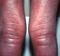 Indurated edematous plaques of hypereosinophilic syndrome on a patient's legs.
 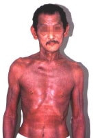 Erythroderma in a affected person with hypereosinophilic syndrome. NextPathophysiology
over the past 2 a long time, tremendous progress has been made in understanding the mechanisms of eosinophil manufacturing, eosinophil programmed cell death (apoptosis), and how eosinophil immunology contributes to both host defenses in opposition to infections and to tissue harm within the host in circumstances of allergic and autoimmune ailments.
the principle stimuli for eosinophil production are interleukin (IL)-5, IL-three, and the granulocyte-macrophage colony-stimulating factor (GM-CSF). These cytokines are also the main alerts that inhibit eosinophil programmed cell dying. consequently, eosinophilia can also be brought on by way of these three eosinophilopoietic cytokines by using increased eosinophil production, by using eosinophil durability, or via a combination of these.[1, 2] as well as, an evolving number of chemotactic cytokines (ie, chemokines) were based as causing eosinophils to migrate from their website of production in the bone marrow into the blood after which into peripheral tissues. These chemokines include eotaxin-1, eotaxin-2, and RANTES (regulated on activation standard T cell expressed and secreted).
Eosinophils are the supply of a lot of cytokines, together with IL-2, IL-3, IL-4, IL-5, IL-7, IL-13, IL-sixteen, tumor necrosis issue-alpha (TNF-alpha), remodeling growth issue-beta (TGF-beta), and RANTES. along with these cytokines, eosinophils are a source of several cationic proteins that additionally make a contribution to the immunologic responses against infectious disease sellers and to tissue damage in allergic and autoimmune ailments. These cationic proteins embrace eosinophil cationic protein (ECP), eosinophil peroxidase (EPO), Charcot-Leyden crystal lysophospholipase, main common protein (MBP), and eosinophil-derived neurotoxin (EDN).
Secondary eosinophilia is a reactive phenomenon pushed via eosinophilopoietic cytokine liberate by using nonmyeloid cells. Eosinophilic differentiation occurs in the bone marrow from myeloid progenitors during the moves of GM-CSF, IL-three, and IL-5. Mature eosinophils are released into the bloodstream where they migrate speedy to peripheral tissues of the bronchial and gastrointestinal mucosa and skin. Their survival is short, until apoptosis is blocked by using cytokines (GM-CSF, IL-three, and IL-5).
Dysregulated manufacturing of those cytokines via more than a few cell populations account for secondary hypereosinophilia reminiscent of seen in nonmyeloid malignancies (eg, Hodgkin lymphoma; transitional cell carcinoma [TCC] of the bladder; adenocarcinomas of the stomach, colon, and uterus; large cell undifferentiated lung carcinomas; and big cell cervical tumors), allergic reactions, parasitic infections, and other stipulations.
main eosinophilias embody both clonal and idiopathic hypereosinophilic syndrome (HES). These disorders have very heterogeneous underlying pathophysiologies, no longer all of that are neatly-defined. they're via definition eosinophilia for longer than 6 months, with out proof of reactive result in and with indicators and signs of organ involvement.[7]
In some neoplastic issues, the hypereosinophilia is part of neoplastic clonal expansion affecting the myeloid lineage. This pathophysiology would describe the eosinophilia in continual myelogenous leukemia (CML), Ph chromosome or BCR-ABL sure; acute myelogenous leukemia (AML), including inv(16), t(sixteen;sixty six)(p13;q22); myeloproliferative ailments; and myelodysplastic syndromes.
a number of hypereosinophilic syndrome (HES) cases exhibit clonal enlargement of strange lymphocytes. Immunophenotypically, they're characterised via aberrant and immature T cells, which show off strange cytokine manufacturing. T-cell receptor gene rearrangements are demonstrated in many. These T cells produce high levels of IL-5, notion to lead to the hypereosinophilia.
Eosinophilia is further labeled as clonal or idiopathic each clinically and pathologically. the world health organization (WHO) proposed criteria to tell apart idiopathic hypereosinophilic syndrome (HES) from chronic eosinophil leukemia (CEL) with predominant eosinophilic differentiation. The analysis of CEL is made if (1) cytogenetic or molecular proof of clonality is existing, (2) an increase in peripheral blasts of greater than 2% or marrow blasts of more than 5% but less than 19% occurs, and (three) other motives are excluded. The underlying chromosomal abnormalities leading to CEL were described in some cases. A deletion on chromosome band 4q12 ensuing in the FIP1L1-PDGFRA (FIR1- like-1-platelet-derived boom factor receptor–alpha) fusion gene reasons an bizarre constitutively activated tyrosine kinase.
These patients display CHIC2 gene deletion in peripheral blood mononuclear cells as a result of this fusion gene. some other fusion gene involving BCR-PDGFRA has been viewed in CML with marked eosinophilia. Mutations involving PDGFRB rearrangements have been described, in addition to FGFR1 (fibroblast increase factor receptor–1) fusions.[2, 8, 9, 10] medical features of eosinophil leukemia outcome from accumulation of leukemic cells in bone marrow, liver, and spleen.[11] Inflammatory mediators from the eosinophils themselves lead to tissue injury to the pericardium, myocardium, endocardium, and fearful device.
ultimately, idiopathic hypereosinophilic syndrome (HES) is the analysis of exclusion in sufferers with marked extended (>6 mo) eosinophilia with multiple organ involvement however without identifiable cytogenetic or molecular abnormalities. Organ harm occurs from liberate of the contents of eosinophilic granules. a few of these cases transform into identifiable entities.
PreviousNextEpidemiologyFrequencyUnited States
In the U.S., in comparison with developing countries, eosinophilia occurs most often as a result of allergic prerequisites, together with drug reactions and atopic bronchial asthma. Parasitic infections are rare.
international
Helminthic infections are the commonest result in of eosinophilia international because of the excessive prevalence of helminthic parasite infections, a couple of of which can be estimated to contain a whole lot of tens of millions of people.
Mortality/Morbidity
patient mortality and morbidity rely on the individual illness related to eosinophilia. Many helminthic infections change into continual ailments that lead to morbidity however no longer mortality. similarly, allergy symptoms and stipulations related to eosinophilia on a regular basis don't lead to mortality. Eosinophilia related to nonmyeloid malignancies does not impact their particular person prognosis or charges of mortality. The mortality and morbidity associated with clonal and idiopathic reasons is related to the level of tissue involvement, damage, or each at diagnosis; how fast therapy is applied; and therapy responsiveness.
Race
No racial predilection exists for eosinophilia, although the incidence of eosinophilia-associated helminthic parasitic infections is extra well-liked in certain geographic areas of the sector.
intercourse
No male or feminine predilection exists in most subtypes of eosinophilia. on the other hand, there is a marked male predominance in clonal problems involving the PDGFRB fusion gene and a small male predominance in clonal disorders of the FGFR1 gene.
Age
folks of all ages will also be plagued by eosinophilia.
PreviousProceed to medical Presentation , Eosinophilia

No comments:
Post a Comment
Note: Only a member of this blog may post a comment.